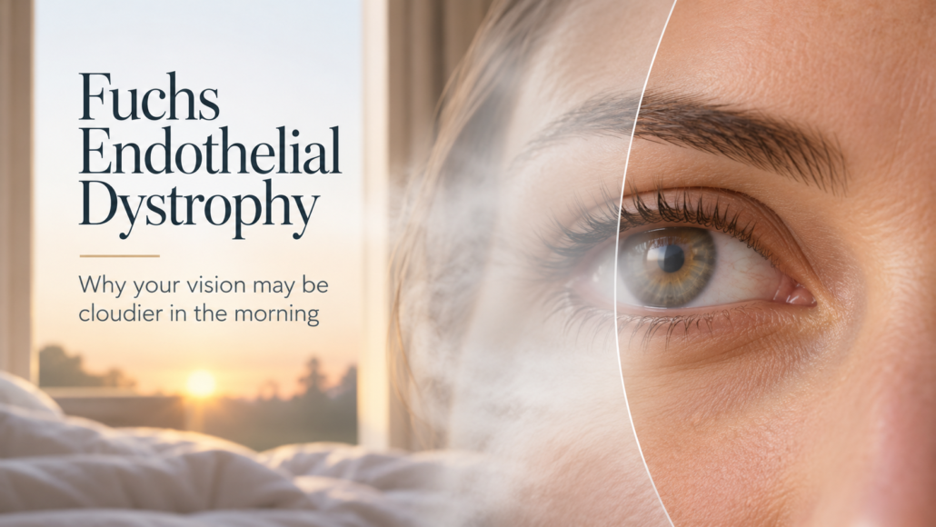
You wake up, open your eyes and everything seems slightly misty.
Your vision is blurred. Lights appear hazy. Reading may take more effort.
Then something curious happens.
As the morning passes, your vision gradually becomes clearer.
It can be tempting to put this down to tired eyes, dry eyes, ageing or simply needing a stronger glasses prescription.
But when vision is consistently cloudier first thing in the morning and improves as the day progresses, there may be another explanation.
It is one of the characteristic patterns associated with Fuchs endothelial dystrophy, a progressive condition affecting the cornea.
The good news is that Fuchs dystrophy can be monitored carefully and, when it begins to significantly affect vision, modern corneal surgery offers highly effective treatment options.
For patients with both Fuchs dystrophy and cataracts, however, treatment planning becomes particularly important.
Mr Vinod Gangwani is a Consultant Ophthalmic Surgeon specialising in corneal disease, cataract surgery and premium lens implants. Having performed more than 9,000 cataract operations, he brings particular expertise to patients whose cataract and corneal health need to be considered together.
First, What Exactly Is Fuchs Endothelial Dystrophy?
To understand Fuchs dystrophy, it helps to think of your cornea as more than simply the transparent window at the front of your eye.
The cornea must remain remarkably clear for light to pass through it properly and create a sharp image.
One of the structures responsible for maintaining that clarity is an extremely thin layer of cells on the inside of the cornea called the corneal endothelium.
These endothelial cells have an important job.
They continually pump excess fluid out of the cornea, helping to keep it transparent.
In Fuchs endothelial dystrophy, these cells gradually deteriorate and reduce in number.
Abnormal deposits known as guttae also develop along Descemet’s membrane, the thin layer supporting the endothelial cells.
During the earlier stages, the remaining endothelial cells can often compensate.
Eventually, however, there may no longer be enough healthy functioning cells to remove fluid effectively.
Fluid begins accumulating within the cornea.
The cornea swells.
And something that should be beautifully transparent starts becoming cloudy.
That is when vision can begin to change.
Why Is Vision Often Worse in the Morning?
This is one of the most interesting characteristics of Fuchs dystrophy.
While you sleep, your eyelids remain closed.
Less evaporation takes place from the surface of the eye, and the cornea naturally retains more fluid overnight.
A healthy endothelium can generally manage this.
An endothelium affected by Fuchs dystrophy may struggle.
The result can be increased corneal swelling by the time you wake, making your vision appear cloudy or misty first thing in the morning.
Once you are awake and your eyes are open, evaporation from the corneal surface increases. In earlier stages of the condition, the cornea may gradually become clearer and vision can improve as the day progresses.
This creates the characteristic pattern:
Cloudy in the morning. Clearer later in the day.
As Fuchs dystrophy advances, however, the cornea may become less able to clear that excess fluid.
The morning blur can take longer to resolve.
Eventually, blurred or hazy vision may remain throughout much of the day.
Other Symptoms You May Notice
Fuchs dystrophy develops gradually, and some people have relatively few symptoms during its earlier stages.
As the condition progresses, you may notice:
- Blurred, cloudy or hazy vision
- Vision that is worse after waking
- Increasing glare from bright lights
- Halos or starbursts around lights
- Greater difficulty driving at night
- Reduced contrast or clarity
- Sensitivity to light
- Fluctuating vision
- Increasing difficulty reading or performing detailed tasks
In more advanced disease, significant corneal swelling can cause tiny fluid-filled blisters to develop on the corneal surface.
If these rupture, the eye can become uncomfortable or painful.
At this stage, Fuchs dystrophy is no longer simply affecting visual quality. The cornea itself is struggling to maintain its normal function.
Fuchs Dystrophy Usually Affects Both Eyes, But Not Always Equally
Fuchs endothelial dystrophy typically affects both eyes.
That does not mean both eyes will progress at exactly the same rate.
One eye may have more significant corneal changes than the other, and symptoms can differ between eyes.
The condition is also generally progressive, but the speed of progression varies considerably.
Some people can have visible signs of Fuchs dystrophy for years without requiring surgery.
Others develop clinically significant corneal swelling and visual deterioration.
This is why discovering Fuchs dystrophy does not automatically mean you need an operation.
The important question is not simply:
“Do I have Fuchs dystrophy?”
It is:
“How much is it affecting the health of my cornea and the quality of my vision?”
What Causes Fuchs Endothelial Dystrophy?
Fuchs dystrophy is associated with a combination of genetic and age-related factors.
Family history can be relevant, although inheritance is not always straightforward.
The condition is generally more commonly recognised later in adulthood and is reported more frequently in women.
What matters clinically is that endothelial cells have very limited ability to regenerate naturally.
When their number falls significantly, the remaining cells have to work harder to maintain corneal clarity.
This is why careful monitoring becomes important as the disease progresses.
How Is Fuchs Dystrophy Diagnosed?
Fuchs dystrophy may sometimes be discovered during a routine eye examination or while someone is being assessed for cataract surgery.
A specialist assessment looks beyond whether you can simply read the letters on an eye chart.
Mr Gangwani may assess several aspects of your cornea and vision depending on your individual circumstances.
Slit-Lamp Examination
A detailed microscopic examination can reveal characteristic corneal guttae, changes in the endothelial layer and signs of corneal swelling.
Corneal Thickness
Corneal swelling can increase the thickness of the cornea.
Measuring corneal thickness, known as pachymetry, can therefore provide useful information when considered alongside the rest of the examination.
Endothelial Cell Assessment
Specialist imaging can help assess the density and appearance of the endothelial cells.
This can be particularly valuable when planning cataract surgery because the procedure itself places additional stress on the corneal endothelium.
Corneal Imaging
Modern corneal imaging can reveal subtle changes and patterns of swelling that may not always be obvious from symptoms alone.
No single number should determine treatment in isolation.
Visual symptoms, corneal appearance, imaging findings, cataract severity and the patient’s individual needs all need to be considered together.
The Important Connection Between Fuchs Dystrophy and Cataracts
This deserves particular attention.
Fuchs dystrophy and cataracts commonly occur in a similar age group.
Unfortunately, both can cause overlapping symptoms.
A cataract can cause:
- Blurred vision
- Glare
- Halos
- Reduced night vision
- Faded colours
- Loss of contrast
Fuchs dystrophy can produce several of the same complaints.
So if somebody has both conditions, which one is actually responsible for their deteriorating vision?
Sometimes it is mainly the cataract.
Sometimes it is mainly the cornea.
Sometimes it is both.
That distinction matters enormously because it can influence which operation should be performed and when.
Can You Have Cataract Surgery If You Have Fuchs Dystrophy?
Yes.
But the health of the cornea needs careful consideration before surgery.
During cataract surgery, ultrasound energy and surgical manipulation can cause some loss of endothelial cells.
For someone with a healthy cornea, this is usually well tolerated.
For somebody whose endothelial reserve has already been reduced by Fuchs dystrophy, the calculation is different.
If the cornea has sufficient reserve and there is little or no significant swelling, cataract surgery alone may be appropriate.
If the Fuchs dystrophy is more advanced, cataract surgery could increase the risk of the cornea becoming persistently swollen afterwards.
This is one reason why having a surgeon experienced in both cataract and corneal disease can be particularly valuable.
The decision is not simply how to remove the cataract.
It is how to achieve the best visual outcome whilst protecting the cornea.
Does Fuchs Dystrophy Affect Lens Implant Choice?
Potentially, yes.
Modern cataract surgery offers a sophisticated range of intraocular lens implants, including lenses designed to reduce dependence on glasses.
But premium technology is not automatically the right technology for every eye.
Fuchs dystrophy can affect contrast sensitivity, visual quality and the optical properties of the cornea. Corneal surgery can also alter measurements relevant to lens power calculations.
This means lens selection needs to consider not only the cataract but also the current condition of the cornea and the possibility of future corneal treatment.
Mr Gangwani’s experience in both premium lens implants and corneal disease allows these factors to be considered together rather than independently.
The objective should be the right lens for the individual eye, not simply the most sophisticated lens available.
What Happens in the Early Stages?
Not everyone with Fuchs dystrophy requires surgery.
Where symptoms are relatively mild, monitoring may be appropriate.
Hypertonic saline drops or ointments may sometimes be used to help draw excess fluid from the cornea and reduce swelling.
These treatments can help manage symptoms.
They do not, however, replace damaged endothelial cells or cure the underlying condition.
Regular assessment allows progression to be monitored and helps determine when the balance may shift from observation to surgical treatment.
When Does Fuchs Dystrophy Need Surgery?
There is no single point at which every patient should undergo surgery.
The decision is individual.
Factors may include:
- How much your vision is affecting daily life
- The degree of corneal swelling
- How long morning blur lasts
- Whether vision remains cloudy throughout the day
- Corneal imaging findings
- Your ability to drive, read or work comfortably
- Whether you also have a cataract
- The health of the other eye
- Your visual expectations and lifestyle
Modern thinking increasingly focuses on the overall functional impact and objective corneal findings, rather than waiting for the cornea to become severely compromised.
The timing matters.
Operating unnecessarily early has little benefit.
Waiting until the cornea is significantly damaged may also make visual rehabilitation more complex.
The goal is to identify the appropriate window for the individual patient.
What Is DMEK?
For patients whose Fuchs dystrophy has progressed sufficiently to require corneal transplantation, one of the principal modern procedures is Descemet Membrane Endothelial Keratoplasty, usually abbreviated to DMEK.
The word “transplant” can understandably sound alarming.
But DMEK is very different from replacing the entire cornea.
Remember, in Fuchs dystrophy the main problem lies in the extremely thin endothelial layer at the back of the cornea.
DMEK takes a remarkably targeted approach.
The damaged Descemet’s membrane and endothelial layer are removed and replaced with a very thin layer of healthy donor tissue containing functioning endothelial cells.
Most of your own cornea remains untouched.
The donor endothelial cells can then resume the essential job of removing excess fluid from the cornea.
As the swelling resolves, the cornea can regain clarity and vision can improve.
Why Has DMEK Changed the Treatment of Fuchs Dystrophy?
Historically, advanced corneal disease often required a full-thickness corneal transplant.
Modern endothelial keratoplasty has fundamentally changed that approach.
Instead of replacing healthy layers of the cornea along with the damaged tissue, DMEK replaces only the layers principally affected by the disease.
For appropriately selected patients, this tissue-specific approach can offer excellent visual rehabilitation while avoiding many of the disadvantages associated with traditional full-thickness transplantation.
DMEK is now an important surgical treatment for visually significant Fuchs endothelial dystrophy.
What Is DSAEK?
Another form of endothelial corneal transplantation is Descemet Stripping Automated Endothelial Keratoplasty, or DSAEK.
Like DMEK, it replaces the diseased endothelial layer.
The difference is that the transplanted tissue in DSAEK includes a thin layer of donor corneal stroma as well as Descemet’s membrane and endothelial cells.
Both techniques have an established role in endothelial corneal disease.
Which approach is appropriate depends on the individual eye, its anatomy, any previous surgery and other clinical considerations.
This is not a decision a patient needs to make from an internet comparison chart.
It is a surgical decision that should follow a detailed examination of the eye.
Cataract Surgery and DMEK: Together or Separately?
This is one of the most nuanced decisions in managing Fuchs dystrophy.
If a patient has both a significant cataract and sufficiently advanced Fuchs dystrophy, there are several possible approaches.
Cataract surgery may be performed first.
DMEK may be performed first.
Or cataract surgery and DMEK may sometimes be performed during the same surgical episode, often referred to as a combined or triple procedure.
There are advantages and disadvantages to each strategy.
Current research has not established one universal approach that is best for every patient. Studies comparing combined and sequential surgery show that both can achieve good outcomes, while factors such as refractive predictability, graft attachment, recovery and the severity of each condition influence decision-making.
This is precisely why treatment should be individualised.
The right question is not:
“Which operation is best?”
It is:
“Which sequence is best for this eye?”
Why Might a DMEK Graft Need a Gas or Air Bubble?
During DMEK surgery, the transplanted tissue needs to attach to the back surface of the patient’s cornea.
An air or gas bubble is placed inside the eye to help support the delicate graft in the correct position while it attaches.
Patients are usually given specific instructions about positioning following surgery.
Occasionally, part of the graft does not attach sufficiently.
If necessary, another air or gas bubble can be inserted to encourage attachment. This is known as rebubbling.
It is a recognised part of DMEK care and does not necessarily mean that the transplant has failed.
Will Fuchs Dystrophy Make Me Blind?
For most patients diagnosed with Fuchs dystrophy, this is understandably one of the biggest fears.
Fuchs dystrophy can significantly impair vision if it progresses and is left untreated.
But modern corneal treatment has transformed its management.
The more useful response to a diagnosis is therefore not panic.
It is appropriate monitoring and timely treatment.
Understanding the severity of your condition and establishing a long-term plan with a corneal specialist can make the diagnosis far less daunting.
Five Signs It May Be Time to Have Your Cornea Checked
Consider seeking a specialist assessment if you are experiencing:
1. Vision that is repeatedly cloudy on waking
Particularly if it takes progressively longer to clear.
2. Increasing glare when driving at night
Headlights may become uncomfortable or surrounded by halos.
3. Vision that fluctuates during the day
Especially when there is a consistent morning-to-evening pattern.
4. A known family history of Fuchs dystrophy
Particularly if you are also developing visual symptoms.
5. Cataracts alongside known corneal guttae or Fuchs dystrophy
A detailed corneal assessment can be particularly important before deciding how cataract surgery should be approached.
Frequently Asked Questions
Is Fuchs endothelial dystrophy hereditary?
Genetic factors can play an important role, and Fuchs dystrophy can occur within families. However, its inheritance and expression can vary. Having a relative with Fuchs dystrophy does not necessarily mean you will develop clinically significant disease.
Does Fuchs dystrophy always affect both eyes?
It usually affects both eyes, although one eye may be more advanced than the other.
Can eye drops cure Fuchs dystrophy?
No. Certain drops or ointments may help manage corneal swelling and symptoms in earlier disease, but they do not restore endothelial cells that have been lost.
Why is my vision worse when I wake up?
The closed-eye environment during sleep reduces evaporation from the corneal surface. In Fuchs dystrophy, the weakened endothelial layer may struggle to remove the additional fluid efficiently, causing increased morning swelling and blurred vision.
Will I definitely need a corneal transplant?
No. Some people remain stable for a considerable period and can be monitored without surgery. The need for surgery depends on disease progression, corneal swelling, visual function and the impact on everyday life.
Can I still have cataract surgery?
Yes, but Fuchs dystrophy should be assessed carefully before cataract surgery. The severity of the corneal disease influences whether cataract surgery alone or a combined or staged approach with endothelial keratoplasty may be more appropriate.
What is the difference between DMEK and a traditional corneal transplant?
DMEK replaces only the very thin inner layers of the cornea affected by endothelial disease. A traditional penetrating keratoplasty replaces the full thickness of the cornea. The appropriate procedure depends on the condition of the individual eye.
How quickly does Fuchs dystrophy progress?
There is considerable variation. Some people progress slowly over many years, while others develop more significant corneal swelling and visual symptoms. Regular assessment helps establish how the condition is behaving in your eyes.
Specialist Assessment with Mr Vinod Gangwani
Fuchs endothelial dystrophy sits at an important intersection between corneal disease and cataract surgery.
That makes specialist assessment particularly valuable.
Mr Vinod Gangwani is a Consultant Ophthalmic Surgeon, FRCS, MRCOphth, specialising in cataract surgery, premium lens implants and corneal disease.
He is a consultant at Ashford and St Peter’s NHS Foundation Trust, where he is Clinical Lead for cataract microsurgery, and has performed more than 9,000 cataract operations.
For patients with Fuchs dystrophy, this combined experience is especially relevant.
The objective is not simply to identify the condition.
It is to understand:
How healthy is the cornea now?
What is causing the visual problem?
Is there also a cataract?
Does anything need to be done now?
And, if surgery is required, what sequence offers the most appropriate strategy for that individual eye?
That is where specialist assessment matters.
Do Not Ignore the Morning Mist
Fuchs Endothelial Dystrophy can progress quietly, and its early signs are sometimes easy to dismiss.
A little morning blur.
More glare from headlights.
Vision that takes longer to clear after waking.
These patterns can provide useful clues about what is happening inside the cornea.
A diagnosis of Fuchs Endothelial Dystrophy does not automatically mean surgery.
It means the cornea deserves closer attention.
With careful monitoring, modern corneal imaging and treatments such as DMEK where appropriate, Fuchs Endothelial Dystrophy can be managed with a treatment strategy tailored to the individual eye.
If you are experiencing persistent morning blur, increasing glare or have already been diagnosed with Fuchs Endothelial Dystrophy, arrange a specialist consultation with Mr Vinod Gangwani for a detailed assessment of your corneal health and treatment options.