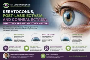

Keratoconus is a degenerative condition causing thinning and a cone-like distortion of the cornea. Anterior lamellar corneal transplant (DALK) is a partial-thickness graft that replaces diseased front corneal layers while leaving the endothelium intact. DALK diminishes the risk of graft rejection, reduces recovery times for many and can restore vision if glasses or rigid lenses fail to assist. For clear context, the following pages detail symptoms, diagnosis, DALK process, risks, recovery, and results.
Understanding Keratoconus
Keratoconus is an ectatic corneal disease in which the clear front part of the eye thins and protrudes in a cone shape. Vision blurs due to irregular astigmatism, glare, and higher-order aberrations that glasses cannot resolve. It can vary significantly both between individuals and even between the two eyes. Early diagnosis with corneal topography, pachymetry, and routine eye care can slow progression and plan timely treatment.
The Condition
Keratoconus means the cornea loses strength and its smooth dome shifts into a cone. The front cornea is worst hit, as the collagen lamellae slip and the frontal layers become steeper and thinner. With chronically active stress, fine fissures and opacification may appear in both the epithelium and Bowman’s layer, and scarring can develop, particularly following hydrops or protracted contact lens wear.
It disturbs the cornea’s normal layered structure, so light scatters rather than hitting the retina cleanly. It is the second leading cause of corneal transplant in young adults globally, and studies show it has now become a more common indication than other factors such as pseudophakic bullous keratopathy. Where transplant access is scarce, scarring can heavily impact life and work.
The Causes
Key components are genetic tendency, chronic eye rubbing, and environmental stress like atopy, dry, dusty air, or ultraviolet light. Family history increases the risk and particular ethnic groups have higher rates, although prevalence rates vary by region and study.
There are associations with allergies, vernal keratoconjunctivitis, connective tissue disorders and Down syndrome. The fundamental tissue defect is pathological collagen and extracellular matrix in the stroma that makes the corneal scaffold weak. What triggers keratoconus is not entirely known. However, these risk factors are well mapped and inform preventative advice, such as rub control.
The Symptoms
You see blur that glasses never seem to correct, increased glare and halos, and rapid changes in prescription numbers. As the cornea thins, irregular astigmatism increases, frequently with additional short-sight. Night vision suffers, with light ghosting and streaks that make driving difficult.
Others experience eye strain and itching, leading to rubs and speed drops. In advanced cases, haze and scarring decrease contrast and can obscure vision in the line of sight.
The Progression
Change often develops gradually from mild to severe across the years, more quickly in the teenage years and early twenties. Care teams monitor it with curvature mapping scans, corneal thickness, and posterior elevation. Small but consistent changes indicate genuine progression. Left untreated, the cornea may scar or swell, which is known as acute hydrops, and surgery, including deep anterior lamellar keratoplasty or other grafts, may be required.
Progress isn’t the same for everyone. Some eyes stabilise with age or after corneal cross-linking, while others keep steepening and require contacts, ring segments, or transplant. Keratoconus is prevalent in the West, frequently runs in families, and given the right treatment, has a good prognosis although transplants have complications such as scarring, rejection, and limits to visual capability. Routine examinations are still essential in order to preserve sight and provide care that matches local access and resources.
Your Keratoconus Treatment Options
– your keratoconus treatment options Treatment ranges from basic visual aids to sophisticated corneal surgery. Choice depends on stage, cornea shape and thickness, scarring, age, rate of change, and daily visual requirements. The aim is twofold: clear vision and slowing or stopping progression. Most don’t require surgery, but plans should remain flexible as keratoconus can change over time.
Non-Surgical
Get Keratoconus Treatment 2023. They clear up blurriness caused by short-sight and astigmatism and are suitable for daily chores once the cornea is still relatively smooth. They are straightforward, low-risk and easily adjustable as prescriptions change.
RGPs provide a clearer image by hiding the irregular corneal surface under a smooth shell. Fit is key. A proficient fitter topographs the cornea and adjusts edge lift and diameter to optimise comfort and vision. As corneal peaks and troughs increase in size, scleral lenses ride high. These large lenses vault the cornea and sit on the whites of the eye, with a salt-water reservoir that smooths optics and may improve comfort for dry or sensitive eyes.
Corneal crosslinking (CXL) is the primary means to slow or stop progression, particularly in younger individuals who generally deteriorate more rapidly. It employs riboflavin drops and controlled UV light to regularise corneal collagen. It does not fix the shape itself, but helps lock the shape in so lenses work longer. Many clinics verify documented change pre-CXL, such as 10 to 20 micrometre thinning or steepening on topography.
Non-surgical treatment suits the majority of mild to moderate cases. Routine checks with topography indicate when to change, and typically the progression will go from glasses to rigid or scleral lenses as the cone becomes steeper.
Surgical
Surgery comes into the picture when lenses no longer provide usable vision or cannot be fitted, or scarring diminishes clarity. Typically, only about 10 to 20 per cent of people will reach this point.
ICRS (intracorneal ring segments) are plastic arcs inserted within the cornea to flatten the steep areas and bring the cornea into a more normal shape. They can reduce irregular astigmatism and assist some patients back into glasses or softer lenses but do not halt progression. CXL is frequently combined for stability.
Transplants are for the more advanced cases. Deep Anterior Lamellar Keratoplasty (DALK) replaces the front layers but preserves your own endothelium, reducing the risk of endothelial rejection and can be judicious if the inner layer is healthy. Penetrating Keratoplasty (PK) is full-thickness and is chosen when scars are deep, the cornea is extremely thin or the Descemet’s/endothelium is damaged. Recovery is slow for both procedures, with vision, on average, taking 6 to 12 months to settle, and stitches are removed in stages. Graft selection is based on thickness maps, scar depth, previous infections and real-life visual objectives.
Deep Dive into DALK Surgery
Deep anterior lamellar keratoplasty (DALK) is a partial-thickness corneal transplantation for diseases that spare the endothelium. It substitutes the anterior corneal layers with donor stroma and retains the patient’s own Descemet’s membrane and endothelium, reducing rejection risk and enhancing long-term graft survival. For keratoconus without endothelial dysfunction, DALK is usually the procedure of choice over full-thickness penetrating keratoplasty (PK) as it lowers complications, increases longevity and preserves endothelial counts.
1. The Procedure
Surgeons excise the diseased epithelium and stroma and insert atop a donor stromal button of comparable size, sutured in with fine nylon stitches. The host Descemet’s membrane and endothelium remain intact, providing a natural barrier against immune attack and protection from full-thickness wounds.
Many cases use the big-bubble technique. A small burst of air is injected into deep stroma to split layers and reveal bare Descemet’s membrane for a smoother interface. When air dissection fails, surgeons resort to layer-by-layer manual debulking.
Specialised tools assist in this work. A Fogla cannula assists air injection, and pneumatic dissection and spatulas direct cleavage at the correct plane. Precision is vital. A clean, even lamellar bed pushes graft clarity, minimises interface haze and ensures the best opportunity for good vision.
2. The Candidacy
Best suited to DALK are patients with keratoconus or analogous stromal disease with an intact endothelium confirmed by the use of specular microscopy.
Patients who have corneal endothelial failure, deep central scarring down to Descemet’s membrane, or had previous hydrops with rupture may be directed to PK.
Pre-operative tests direct the phonecall. Corneal topography maps cone shape, pachymetry checks thickness, and tomography flags risk of perforation. DALK can be done as a first graft or as a regraft when the endothelium is still good.
3. The Technology
Femtosecond laser‑assisted DALK can incise exact trephination depths and zig‑zag edges, improving adaptation and sometimes hastening suture removal. Intraoperative OCT provides real-time depth perception cues, reducing perforation risk and assisting in determining when to cease dissection. Eye banks provide sterile, quality-checked donor corneas with measured stromal thickness and endothelial counts. Novel cannulas, guarded dissectors and more benign viscoelastics are designed to improve the safety and speed of surgery.
4. The Comparison
Compared with PK, DALK has a far lower risk of endothelial rejection and less endothelial cell loss. Recovery may be smoother because the globe is never opened full thickness and trauma risk is reduced. High astigmatism is still frequent following DALK, so suture management and selective relaxing incisions are important.
- Visual acuity: DALK is approximately equal to PK when the interface is clear.
- Complications: DALK fewer immune rejections; PK higher endothelial failure
- Long‑term health: DALK preserves native endothelium better than PK
5. The Success Rate
Large series show strong survival in keratoconus. The 10-year DALK survival rate is around 94% compared to 72% for PK. Quicker, steadier visual rehabilitation is the norm with a smooth interface and well-handled sutures. DALK’s reduced endothelial cell loss probably underpins improved long-term clarity. Outside keratoconus, DALK aids in stromal dystrophies and scars with intact endothelium, enhancing acuity and alleviating glare and ghosting.
The DALK Patient Journey
A clear path helps patients prepare, measure choices, and pace recovery. DALK replaces the diseased front cornea but leaves the inner layer intact for safer grafts and gradual vision improvements over months. A standard journey includes multiple teams, intricate tissue logistics, a two-hour operation, and aftercare that extends to a year or more.
- Diagnosis and optimisation: Comprehensive eye exams, topography, tomography, and pachymetry confirm keratoconus stage and graft suitability. Talk about alternatives like cross-linking or special lenses.
- Surgical planning and consent: The surgeon explains risks, benefits, anaesthesia choices, costs, timelines, and time off work (often 1 to 2 weeks). Eye bank coordination secures appropriate donor tissue.
- Operation day: Expect local or topical anaesthesia, two-hour theatre time, and half-day in hospital. From the corneal surgeon to the anaesthetist, scrub team and recovery nurses, everyone is involved.
- Early recovery (days–weeks): Blurred vision for one to two weeks is common, with grittiness and light sensitivity. Instill drops, shield the eye, and pace the day.
- Ongoing recovery (months): Vision improves slowly over six to twelve months, sometimes up to twenty-four months, with glasses or contact lenses for best results. Most patients, around seventy-five per cent, attain aided driving vision.
- Long-term stewardship: Regular checks monitor graft clarity, suture status and rare complications.
Before Surgery
Expect a full work-up: refraction, slit-lamp exam, corneal maps, and thickness scans. This establishes a baseline and directs graft size.
Stop soft lenses for a week or rigid lenses for two to four weeks, as suggested, so the cornea can settle for accurate measurements.
Consent covers common issues such as slow visual recovery and the need for glasses, as well as rare but serious risks including endophthalmitis, severe bleeding, retinal detachment, and marked inflammation. Eye bank teams screen donor tissue for quality and disease. Scheduling aligns the surgeon, theatre, and tissue delivery.
During Surgery
Anaesthetising usually involves topical drops along with sedation or a local block. General anaesthesia is available if necessary.
The surgeon lamellar dissects down to Descemet’s, places the donor lamella, then sutures with fine nylon. Chamber stability is key throughout. Judicious viscoelastic use, controlled pressure and careful suturing prevent perforation and maintain centration and low astigmatism.
After Surgery
You go home the same day with drops and a shield. Vision is hazy at first. Light bothers some. Grit-like feel is normal.
- Medication: Steroid and antibiotic drops as prescribed, tapering over months. Lubricants for comfort.
- Protection: Night shield for at least one week. No eye rubbing. Keep soap and makeup away from the eye for two weeks.
- Activities: No swimming or gardening for four weeks. Avoid heavy lifts.
- Work: Plan one to two weeks off, based on job demands and surgeon advice.
- Follow-up: Early visits check pressure, graft clarity, suture tension, and signs of infection or inflammation. Later visits guide suture removal and optical correction. Urgent review with sudden pain, drop in vision, or new floaters.
Navigating Risks and Outcomes
Keratoconus thins and distorts the cornea. DALK replaces diseased stroma but leaves the patient’s own endothelium, with faster healing and a lower rejection risk than full-thickness transplant (PK).
Potential Complications
- Intraoperative: microperforation of Descemet’s membrane, incomplete stromal dissection, loss of the big bubble, interface bleeding, and high intraocular pressure spikes from overfilling the anterior chamber.
- Postoperative complications include graft detachment or partial apposition, interface haze or scarring, irregular astigmatism, epithelial defects, infection from bacterial, fungal, or herpetic sources, steroid-induced rise in intraocular pressure, and suture-related problems.
- Rare events include persistent interface fluid, endothelial trauma converting to PK, and graft failure requiring repeat keratoplasty.
Pre-op control of surface disease, quieting any active inflammation or infection, and evaluating cataract is key. Treat first if it clouds vision. Maintaining safe IOP intra and post-op helps keep the interface clear, the optic stable, and the nerve healthy.
In patients where DALK reaches Dua’s layer, later phacoemulsification is possible with less risk to the endothelium. This is important if cataract surgery is delayed until the graft has stabilised.
Graft Rejection
DALK sharply reduces the risk of endothelial rejection compared with PK because the host endothelium remains. This doesn’t render rejection impossible; stromal or epithelial immune episodes can still ensue.
Look out for: Redness, ache or pain, sensitivity to light, and a decrease in vision. A good new haze at the interface or new KPs along the graft edge can be early signs.
Timely Treatment is Most Effective. Start or escalate topical steroids, add lubricants, and manage lids and allergy triggers. In obstinate cases, brief runs of systemic immunosuppression or steroid-sparing agents do the trick. Close follow-up matters most in the first year, then lifelong at intervals.
Long-Term Vision
When the interface is clear and the cornea remains clear, the quality of the vision is usually stable for years. Most patients obtain good functional vision with optimal results once sutures have been completely taken out and the cornea stabilises.
We always see through glass clearer. Many still opt for rigid gas-permeable or scleral lenses to neutralise irregular astigmatism. That is a choice based on comfort, practicality and price.
Sustained transparency with consistent medication and stable IOP is the norm. Large series demonstrate graft survival in keratoconus of 96.7% at 10 years and 95.6% at 20 years. Survival in stromal disease is greater following DALK than PK, with significantly improved 50-year outcomes. However, for very young children (less than 5 years), risks of failure are comparable. At 5 years, differences in visual acuity and corneal thickness between DALK and PK often converge, but DALK still reduces rejection risk and facilitates safer cataract surgery in the future.
| Outcome metric | Typical trajectory after DALK | Quality of life impact |
| Best-corrected VA | Improves over 6–18 months, then stable | Easier reading, driving in good light |
| Corneal clarity | High with low rejection risk | Less glare, sharper contrast |
| Spectacles/contacts | Common for fine-tuning | Better work and study performance |
| Graft longevity | Strong long-term survival | Fewer re-operations |
A Surgeon’s Perspective on DALK
From the theatre table, DALK straddles older full-thickness grafts and newer selective techniques. It preserves the patient’s endothelium, reduces immune risk and still requires delicate hands. Excellent DALK relies on careful planning, depth control and tranquil problem-solving when the cornea surprises you.
Why DALK?
DALK is selective by design. We excise the infected anterior stroma and leave the host endothelium and Descemet’s intact, typically intact in keratoconus. That simple act of preservation changes everything about the risk profile and the long-term care. It’s one of the reasons we rank DALK first for keratoconus without endothelial disease.
Rejection risk falls because the graft dispenses with donor endothelium, the most antigenic layer. Graft survival is longer, with reduced late failures due to endothelial loss. In clinic, this often means less steroid exposure and fewer pressure spikes.
Visual recovery is steadier with a clean interface and Descemet’s intact. Complications, such as wound leaks and expulsive events, are less common than with penetrating keratoplasty. Rehab isn’t fast. Clear vision may take over 12 months, with steps on the way: new glasses, specialty contact lenses, staged suture removal or adjustment, and sometimes keratorefractive tweaks for irregular astigmatism. Some will subsequently require cataract or glaucoma surgery, arranged with the graft in mind.
The technique matches the broader shift to tissue-sparing corneal surgery. Choose what replaces and preserve that which still serves the eye and contain systemic risk. That’s a useful, patient-centred shift.
Future Innovations
Regenerative tools will likely blend with DALK. Stromal cell therapy will strengthen host tissue and bioengineered anterior stroma will ease donor gaps and standardise quality.
Imaging will control depth in real time. Pentacam findings already support big-bubble planning. Research indicates that a 90% intended trephination depth enhances reliable bubble formation. Add intraoperative OCT and cleverer surgical navigation, and nailing the right plane becomes like clockwork rather than artistry.
Eye banks will refine donor handling for anterior lamella by providing better storage media for stromal clarity, pre-cut anterior buttons, and tighter thickness specifications. That consistency cuts theatre time and reduces interface opacity.
With safer instruments, indications can expand. Earlier keratoconus with scarring, post-infective anterior opacities, and even some trauma would benefit. Take-up should increase as training becomes better, but results will remain reliant on technique and case selection.
Attention to technical detail matters. Many surgeons decimate the outer two-thirds of the stroma in keratoconus to insert a 27-gauge needle or cannula deep for air injection or viscodissection. Trephination depth is checked with a paracentesis blade, and big-bubble attempts begin just shy of 90% recipient depth. If microperforations affect less than 25% of the cornea, we usually go using air or gas tamponade to re-appose DSAEK. Suture strategy differs from PK: in DALK, pass sutures about 50% depth in the donor and 90% in the host, improving Descemet’s apposition; in PK it is 90% in both. Some prefer adjustable speculums to eliminate excess globe pressure, which decreases the likelihood of perforation and maintains a steady field.
Conclusion
Keratoconus can certainly feel like a long road. Clear steps rest on the map. Glasses and lenses assist many. Cross Linking can hold shape. DALK preserves your native cells and can provide sharp clarity. There are reduced graft risks compared to a full graft. Wound healing takes time, but most observe stable improvements by month 3 to 6.
A short story. One young chef traded ghosted street lights and sharp knife work for DALK. Not flawless on day one. Far better by week eight. Back on the line by month four.
To consider next steps, consult with a cornea surgeon who performs DALK regularly. Ask for results, rates and waits. Bring your ambitions and daily requirements. Book that consult and get a clear plan.