Understanding Conditions That Affect the Strength and Shape of Your Cornea
The cornea is one of the most important structures of the eye. It plays a crucial role in focusing light and allowing you to see clearly. When the cornea becomes weakened and changes shape, it can affect vision quality and, in some cases, lead to progressive sight problems.
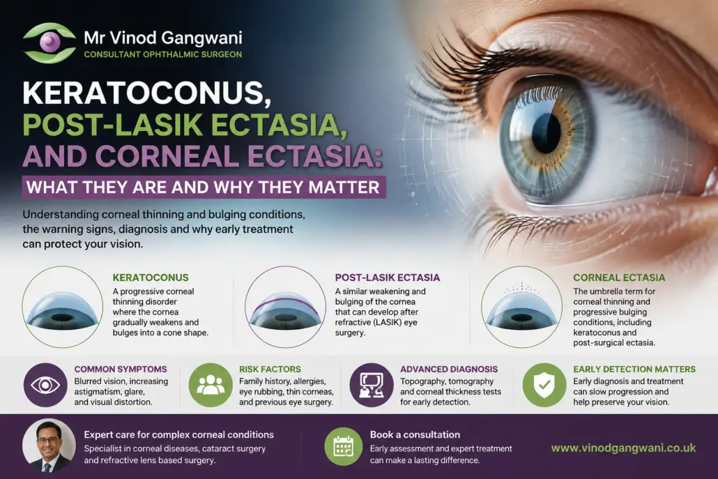
Conditions such as keratoconus, post-LASIK ectasia, and corneal ectasia all involve changes in the strength and structure of the cornea. Although they are related, they are not exactly the same condition.
Keratoconus is a naturally occurring progressive condition where the cornea gradually becomes thinner and starts to bulge forward.
Post-LASIK ectasia is a similar weakening and bulging of the cornea that develops after refractive laser eye surgery in some patients.
Corneal ectasia is the broader medical term used to describe corneal thinning and progressive bulging conditions, including keratoconus and post-surgical ectasia.
Early diagnosis and specialist management are important because treatments are most effective when these conditions are identified before significant progression occurs.
Mr Vinod Gangwani is an experienced ophthalmic surgeon specialising in corneal diseases, cataract surgery and advanced lens-based procedures. Having gained extensive experience at Moorfields Eye Hospital, he provides specialist corneal care for patients with both routine and complex corneal conditions.
What Is the Cornea and Why Does It Matter?
The cornea is the clear, dome-shaped surface at the front of the eye that covers the iris and pupil.
Its main functions include:
- Protecting the eye from dust, bacteria and injury
- Controlling how light enters the eye
- Providing much of the eye’s focusing power
A healthy cornea has a smooth, regular shape that allows light to pass through accurately onto the retina.
When the cornea becomes weakened, it may lose its normal shape. Instead of maintaining a smooth curve, it can become thinner and develop a cone-like or irregular bulging shape.
This can cause:
- Blurred vision
- Distorted images
- Increasing levels of astigmatism
- Difficulty achieving clear vision with glasses or contact lenses
What Is Keratoconus?
Keratoconus is a progressive corneal thinning disorder where the cornea gradually becomes weaker and changes shape.
The word “keratoconus” comes from:
- Kerato, meaning cornea
- Conus, meaning cone
As the condition progresses, the normally rounded cornea becomes increasingly cone-shaped.
This irregular shape affects how light is focused inside the eye, leading to changes in vision.
Keratoconus usually develops during the teenage years or early adulthood, although it can occur at any age.
The progression varies between individuals. Some people experience slow changes over many years, while others may have more rapid progression.
What Causes Keratoconus?
The exact cause of keratoconus is not fully understood. It is believed to involve a combination of genetic and environmental factors.
Research suggests that factors contributing to keratoconus may include:
- Genetic susceptibility
- Weakening of collagen fibres within the cornea
- Eye rubbing
- Allergic eye conditions
The cornea is made up of collagen fibres that provide strength and structure. In keratoconus, these fibres may become weaker, allowing the cornea to gradually thin and bulge.
What Is Post-LASIK Ectasia?
Post-LASIK ectasia is a rare complication that can occur after laser eye surgery such as LASIK.
LASIK works by reshaping the cornea to correct vision problems such as:
- Short-sightedness (myopia)
- Long-sightedness (hyperopia)
- Astigmatism
During LASIK surgery, a controlled amount of corneal tissue is removed to alter the shape of the cornea.
In some patients, the cornea may not have sufficient structural strength after surgery. Over time, this can lead to progressive thinning and forward bulging, similar to keratoconus.
This condition is known as post-LASIK ectasia.
Who Is More At Risk of Post-LASIK Ectasia?
Modern laser eye surgery screening has significantly reduced the risk of post-LASIK ectasia.
However, certain factors may increase the likelihood of developing this condition, including:
- Previously undiagnosed keratoconus
- Abnormal corneal shape before surgery
- Thin corneal thickness
- Family history of corneal disorders
- Certain corneal biomechanical characteristics
This is why detailed pre-operative assessment is essential before considering laser vision correction.
Advanced corneal imaging allows surgeons to assess the shape, thickness and strength of the cornea before recommending treatment.
What Is Corneal Ectasia?
Corneal ectasia is the umbrella term used to describe conditions where the cornea becomes progressively thin and develops an abnormal outward bulging shape.
Keratoconus and post-LASIK ectasia are both forms of corneal ectasia.
Other examples include:
- Pellucid marginal degeneration
- Post-surgical corneal weakening
- Other progressive thinning disorders
Although these conditions have different causes, they share similar features:
- Reduced corneal strength
- Progressive thinning
- Irregular corneal shape
- Visual distortion
Common Symptoms and Warning Signs
Symptoms can vary depending on how advanced the condition is.
Early signs may be subtle and can sometimes be mistaken for normal changes in eyesight.
Common symptoms include:
Increasing Blurred Vision
Patients may notice that their vision becomes less clear despite wearing glasses or contact lenses.
Increasing Astigmatism
A changing glasses prescription, particularly increasing astigmatism, can sometimes indicate changes in corneal shape.
Difficulty Seeing Clearly at Night
Some patients experience:
- Glare
- Halos around lights
- Reduced night vision
Frequent Changes in Prescription
Needing regular updates to glasses or contact lenses may be a warning sign.
Eye Strain or Visual Distortion
Some patients describe:
- Difficulty focusing
- Distorted images
- Reduced visual quality
If you notice ongoing changes in your vision, an assessment by an eye specialist is recommended.
Risk Factors: Who Is More Vulnerable?
Certain individuals have a higher likelihood of developing corneal ectasia.
Risk factors include:
Family History
Keratoconus can sometimes run in families.
Age
Keratoconus often begins during adolescence or early adulthood.
Eye Rubbing
Frequent or vigorous eye rubbing is associated with an increased risk, particularly in people who already have a genetic tendency.
Allergic Eye Conditions
Conditions that cause itching and irritation can lead to increased eye rubbing.
Examples include:
- Hay fever-related eye irritation
- Allergic conjunctivitis
- Chronic eye allergies
Previous Eye Surgery
Patients who have undergone corneal refractive surgery require appropriate screening and monitoring.
How Do Doctors Diagnose Keratoconus and Corneal Ectasia?
Diagnosis involves detailed examination of the cornea using advanced imaging technology.
A routine eye test alone may not always detect early disease.
Specialist investigations may include:
Corneal Topography
Corneal topography creates a detailed map of the corneal surface.
It helps doctors identify:
- Irregular corneal shape
- Areas of steepening
- Early signs of keratoconus
Corneal Tomography
Corneal tomography provides detailed information about:
- Front and back surfaces of the cornea
- Corneal thickness
- Structural changes
This is particularly important for detecting early ectasia.
Corneal Thickness Measurement
Known as pachymetry, this measures the thickness of the cornea.
A thinner cornea may require closer monitoring.
Vision Assessment
Doctors assess:
- Visual acuity
- Refraction changes
- Astigmatism patterns
Together, these tests help determine whether the condition is stable or progressing.
Why Early Detection Matters
Early diagnosis of corneal ectasia is important because treatment options are more effective before significant progression occurs.
If detected early, specialists can monitor changes and consider treatments designed to strengthen and stabilise the cornea.
One important treatment option is:
Corneal Cross-Linking
Corneal cross-linking is a procedure designed to strengthen the corneal collagen fibres.
The aim is to:
- Slow or stop progression
- Preserve existing vision
- Reduce the risk of further corneal weakening
The suitability of treatment depends on factors including:
- Age
- Disease progression
- Corneal thickness
- Overall eye health
Can Keratoconus and Corneal Ectasia Be Treated?
Treatment depends on the severity and progression of the condition.
Options may include:
Monitoring
For mild and stable cases, regular specialist monitoring may be appropriate.
Glasses
In early stages, glasses may provide clear vision.
Specialist Contact Lenses
Rigid gas permeable or scleral contact lenses may improve vision by creating a smoother optical surface.
Corneal Cross-Linking
Used to strengthen the cornea and slow progression.
Advanced Surgical Options
In severe cases where vision cannot be corrected effectively, procedures such as corneal transplantation may be considered.
Specialist Corneal Care With Mr Vinod Gangwani
Keratoconus, post-LASIK ectasia and other corneal ectasia conditions require specialist assessment because early changes can be difficult to detect without advanced imaging.
Mr Vinod Gangwani is an experienced ophthalmic surgeon with specialist expertise in corneal diseases, cataract surgery and refractive lens-based surgery.
Having trained and worked at Moorfields Eye Hospital, he provides tertiary corneal expertise for patients with routine and complex corneal problems.
If you are experiencing changing vision, increasing astigmatism or concerns following laser eye surgery, a specialist corneal assessment can help identify the cause and guide the most appropriate treatment options.