
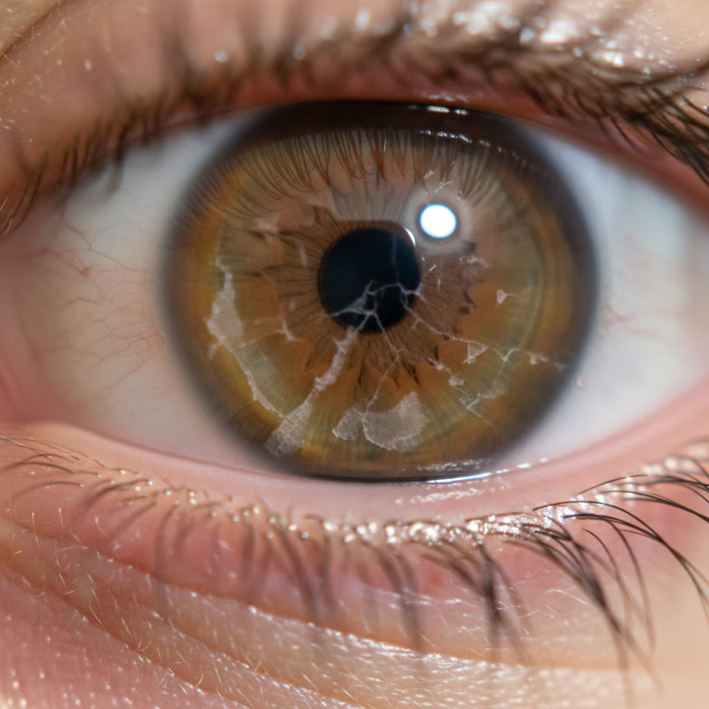
Most commonly, recurrent corneal erosions are caused by prior eye trauma, corneal dystrophies such as epithelial basement membrane dystrophy, and dry eye which weakens surface adhesion. What leads to recurrent corneal erosions? Poor lid fit, night-time lagophthalmos, and wearing contact lenses increase the risk. Diabetes and meibomian gland dysfunction contribute too. To understand triggers, symptoms, and treatment alternatives, the following sections explore causation, diagnosis, and treatment options.
What Causes Recurrent Corneal Erosions?
Recurrent corneal erosions are repeated occurrences in which the corneal epithelium breaks down, exposing the underlying tissue and prompting extreme pain, light sensitivity, and tearing. The fundamental defect is reduced adhesion of the epithelium to its basement membrane. Triggers include prior trauma and corneal disease, with a higher risk in dry eye, corneal dystrophies and following surgery.
1. Previous Injury
The scene may be set by an earlier corneal graze from a fingernail, paper edge, leaf or paw. Even though the initial wound may appear healed, the epithelium can attach poorly to the basement membrane.
When the healing process is incomplete, little patches of loose epithelium develop. When lids peel off a dry surface on waking, those patches can be sheared. Even slight jolts, such as contact lens wear, eye rubbing or head trauma, can trigger the cycle again.
These weaknesses frequently reside in the same location, meaning the same eye flares up again. Lacerations, foreign bodies or a scratch beneath a contact lens all fall into this category.
2. Corneal Dystrophy
Inherited corneal dystrophies make the “glue” between layers less reliable. Epithelial basement membrane dystrophy (map–dot–fingerprint), granular dystrophy, lattice corneal dystrophy, and macular corneal dystrophy are notorious offenders.
The basement membrane can be thick, wavy, or misshaped and so epithelial cells fail to anchor. That defect induces recurrent corneal erosion syndrome and frequently affects both eyes. For a lot of people, specialist treatment is required as conservative measures alone may not suffice.
3. Faulty Healing
Healing can go awry. Cells may migrate incorrectly, deposit poor adhesion and leave holes.
Increased matrix metalloproteinase activity can degrade the very proteins that adhere the cells. Chronic inflammation, certain topical medications and systemic diseases such as diabetes or rheumatoid arthritis contribute to the lag. Each erosion injures the surface again, so recovery becomes slower and relapse more likely.
4. Dry Eye Disease
Too little tear film yields more friction with every blink, so the epithelium tears. Nerve signals desensitise in dry eye, so micro-injuries are ignored and heal slowly.
Meibomian gland dysfunction and ocular surface inflammation destabilise the tear film. Treating dry eye is pivotal to long-term control because a stable tear film protects the epithelium day and night.
5. Other Conditions
Eyelid issues such as lagophthalmos, incomplete closure, and nocturnal exposure dry out the cornea and give rise to exposure keratopathy. Systemic disease matters; diabetes, ocular rosacea, and poor corneal innervation from diabetes or herpes zoster reduce healing. Previous surgery, whether cataract or refractive, can change curvature or the epithelial-stromal interface, while structural alterations such as thinning or abnormal shape aggravate shear forces. Infection, whether bacterial or viral, can provoke or extend defects. For some, the cause remains unknown and is termed idiopathic.
Recognising the Symptoms
Recurrent corneal erosion has a clear and sharp way of announcing itself. Knowing the pattern helps you identify what is normal healing pain versus what indicates the corneal surface not binding well. Symptoms typically flare in episodes that wax and wane over weeks or months. They might wake you up at night, and they can be debilitating enough to interfere with everyday activities, from reading to screen time.
- Sudden, stabbing eye pain on waking, usually as if you’ve been cut or scratched.
- Tearing that does not settle promptly
- Vision blurs then clears, only to fog again, as the eye waters.
- Light sensitivity (photophobia), with glare from lamps or daylight
Most people complain of a foreign body sensation, as if a grain of sand is under the lid. This is usual with flares and can persist as a nagging pain when the more acute pain subsides. The pain usually begins on waking, when the eyelid lifts and tugs on the delicate corneal surface. It can attack randomly during the day, but night and early morning are classic. Peering with one eye closed, walking cautiously in dark rooms or dodging fans and draughts is a dead giveaway that the cornea is inflamed and raw.
Redness is common, not invariably intense. The eye may water enough for tears to run down the cheek, and the temptation to rub is irresistible. Avoid rubbing as it can exacerbate the epithelial slip. On waking, the lids may feel glued together, and it can be difficult to open them. Others discover how to mitigate the first blink by moistening the eye before opening with a lubricating drop kept by the bedside. If that little step sharpens pain, it corroborates the notion of a brittle surface.
Look out for vision fluctuating from sharp to blurry within minutes. Variability of vision, with ghosting or halos about lights, frequently accompanies chronic discomfort. Light sensitivity can be brutal. A darkened room, sunglasses or a baseball cap will serve you well until the attack subsides. Pain varies from a mild sting to stabbing bursts. How intense and frequent it is differs for everyone. Track the pattern: time of day, triggers like dry air or screen strain, and how long each episode lasts. If attacks are recurrent or interfere with work, driving, or sleep, have an eye examination. Recurrent corneal erosion is treatable, and prompt treatment can stabilise the surface.
The Diagnostic Journey
Diagnosis adds symptoms to findings and experience. The goal is to catalogue triggers of recurrent corneal erosions (RCE) and select treatment that matches the pattern, not merely the attack.
Clinical Examination
History first. You’d be asked questions about previous eye injuries (even little scratches from a fingernail), surgery, contact lens use, and work or sport risk factors. They mention family history of corneal dystrophy and medical problems such as diabetes mellitus. They often report pain on waking, tearing, glare and blur that subsides through the day. They note frequency, triggers like dry rooms or a restless night, and how long attacks last. This registers whether the case is post-traumatic, dystrophy-related or mixed.
Fluorescein dye reveals breaks in the epithelium and traces the margins of loose tissue. A large crescent stain under the upper lid suggests an earlier scraping. Microspecks indicate dry eye or blepharitis.
At the slit lamp, the clinician searches for microcysts, map-dot lines or fingerprint patterns of epithelial basement membrane dystrophy. Infection, inflammation, Salzmann’s nodules, band keratopathy and early scarring are excluded. They check lids for incomplete closure, lash trauma and meibomian disease. Tear film break-up time and corneal sensitivity identify dry eye or nerve problems that delay healing. These hints steer gentle measures such as evening drops and eyelid handling or more focused interventions like bandage lenses.
| Finding | What it suggests | Why it matters for treatment |
| Morning pain with fluorescein-positive defect | Classic RCES after trauma | Night ointment, hypertonic drops, bandage lens |
| Map-dot changes and microcysts | Epithelial dystrophy | Debridement with alcohol delamination, anterior stromal puncture |
| Reduced tear break-up time | Evaporative dry eye | Lid hygiene, liposomal sprays, tear substitutes |
| Nerve loss on testing | Neurotrophic surface | Avoid toxic drops, consider serum tears |
| Salzmann’s nodules/band keratopathy | Surface irregularity | Treat underlying lesion to stop recurrences |
Advanced Imaging
Advanced imaging is usually not required, as most cases can be diagnosed by clinical examination..
Modern Treatment Options
Treatment correlates with how often erosions return and how much pain or blurring they create. Care begins with medical therapies and advances to procedures only when episodes persist in the face of months of diligent care.
- Mild or infrequent episodes: medical therapies first-line
- Frequent, painful, or non-healing cases require intensified medical care and a bandage lens.
- Recalcitrant or severe erosions: consider surgical procedures
Medical Therapies
Lubricants set the foundation. Daytime preservative-free artificial tears reduce friction. Stronger gels aid during screen use or dry air. At night, ointments protect with a soft seal. Several clinicians add hypertonic (salt) ointment 5% at bedtime for weeks to months, as it pulls fluid from the cornea and can shorten morning pain.
When the epithelium is open, a topical antibiotic drop or ointment reduces the risk of infection. The choice is easy, wide-ranging, short course, and stops once the surface seals.
Hypertonic saline drops or ointment reduce corneal swelling and enhance epithelial adhesion. This is unflashy, slow build care rather than a fast track and typically lasts 8 to 12 weeks. Those who wake with symptoms tend to take longer.
A bandage contact lens (BCL) provides immediate comfort and protects repair cells from the lid. It stabilises the surface after debridement or AM. Fit is near enough, with routine reviews and antibiotic cover. Some wear a BCL for two to six weeks to get through the high-risk window.
Surgical Procedures
Alcohol delamination with Superficial keratectomy removes loose epithelium and smooths the basement membrane so new cells can anchor. It is easy, done under the slit-lamp or in theatre, and goes well with a BCL and hypertonic ointment at night!
Phototherapeutic keratectomy (PTK) uses an excimer laser to smooth microridges and basement irregularity. It enhances adhesion and has an 80-90% success rate in preventing recurrences. Surgeons map depth meticulously to prevent over-treatment while supplementing it with months of lubricants.
Anterior stromal puncture leaves small, superficial holes in Bowman’s layer to provoke firmer bonding. It is well suited to peripheral lesions as central marks can induce glare. Some centres use a short-pulsed Nd:YAG laser to create precise micro-injuries.
Amniotic membrane options, cryopreserved or dehydrated, bring anti-inflammatory and pro-healing cues. A membrane can remain for 3 to 5 days, then a BCL retains the repair for 2 to 6 weeks. Amniotic membrane debridement has lower recurrence than debridement with BCL only. In stubborn cases, membrane placement may be paired with a short-pulsed Nd:YAG step to improve results. Serum eye drops from donors are an alternative when nerves are not viable or defects persist.
Can This Condition Cause Blindness?
While recurrent corneal erosion (RCE) can lead to partial vision loss, it does not typically produce complete blindness. The majority have painful flares with blurred vision that resolve frequently within hours to days, then see well in between attacks. Even so, if neglected or inappropriately managed, it can damage vision, so the stakes are real.
RCE isn’t directly associated with a risk of blindness and overall, the risk is low. It’s characterised by intense pain on waking, tearing, photosensitivity and intermittent blurred vision. These vision changes are typically temporary as the surface layer, the epithelium, lifts, then heals. Many readers will know the cycle: a minor scratch from a fingernail or past infection, then months of on-off pain. That pain can be excruciating and exhausting, but permanent blindness is rare.
No, the worry isn’t the erosion itself but what comes after if the healing is sluggish or the surface remains rippled. Repeated breakdown can invite microbes in, resulting in infectious keratitis. That can lead to a corneal ulcer. In rare instances, deep ulcers or perforation can develop, and these can endanger vision if untreated promptly. Scarring may occur after bad flares or infection. This can make vision blurry or cause glare, but is not blindness. Think of it like frost on glass: not blacked out, but less clear.
Unwanted astigmatism can occur if the cornea heals unevenly. That can lead to ghosting, night-time glare, haloes round lights and less contrast. People talk about difficulty with screens or driving at night. These can be mild to annoying and can vary from one day to the next, which is exasperating even when chart acuity appears good. Chronic eye pain alters the way you live. Sleep suffers as episodes hit you on waking. Work stalls when your eyes water and sting you through the morning. Quality of life is as much of an issue as line-of-sight vision.
Fast diagnosis and consistent treatment decrease the risk of severe trouble. Lubricants, hypertonic saline at night and lid hygiene keep the epithelium stuck down. Treat meibomian gland dysfunction if present, as oil layer problems delay healing. For obstinate cases, bandage contact lenses, anterior stromal puncture, Alcohol delamination or phototherapeutic keratectomy can reduce recurrences and lower scarring risk. If pain spikes, light hurts or vision drops worse than normal, get urgent care to rule out an ulcer. Most, with the right plan, avoid lasting vision loss.
My Perspective on Prevention
Prevention starts the day the first scratch occurs. Timely and consistent management reduces the chance that an occasional graze becomes a long, painful history of recurrent corneal erosion, in which pain flares with every blink, nights are fraught, and recovery stretches into months or years.
Early treatment of the initial injury is important because loose epithelial cells can heal in a weak bond. A fingernail or twig usually initiates. Twenty-four hours a day, seven days a week contact lens wear can prepare the cornea for trouble too. I encourage people to get treatment immediately after any acute scrape. A doctor could check for loose edges, smooth the surface and initiate a regime with preservative-free lubricants, a bandage contact lens if necessary, or even an early measure like debridement to strip out abnormal tissue. This initial window is when we can intervene in the spiral before it hardens.
Daily lubrication is where it’s at. A truly conditioned lubricant saturates the surface so lids slide, not catch. Regular drops throughout the day keep the tear film stable. At night, a thick ointment – be liberal! – prevents the lid from adhering to the cornea on awakening. A lot of them talk about this sharp tearing pain with the first blink after sleep. The goal is to prevent that moment ever occurring. For high-risk jobs or sports, slip on wraparound eye protection to prevent fresh micro-traumas. Are you wearing lenses? Steer clear of overnight wear, switch to daily disposables or give your cornea a rest while it strengthens its bonds.
Eyelid care supports the oil layer of tears. Warm compresses and gentle lid massage get your meibomian glands moving and slow evaporation. Wipe lid margins daily with a mild non-irritant wipe. Treat dry eye with frequent lubricants, omega-3 in the diet if appropriate, and check medicines that may dry eyes out. When drops and lid care are not enough, targeted procedures can prevent relapse. Careful mechanical removal of loose epithelium by hand or phototherapeutic keratectomy (PTK) can polish the basement membrane. These are designed to provide the new epithelium with a better “bed” to anchor. Recovery can be slow, with the complete healing arc extending over years, so pacing and patience assist.
- Build your plan
- Map triggers: morning pain, air-con exposure, long screens.
- Daily: preservative-free drops every two to three hours; dense nocturnal salve.
- Lids: warm compress 5–10 minutes, cleanse once daily.
- Protection: wraparound glasses for wind, dust, sport.
- Lenses: no overnight wear; consider a lens holiday.
- Home: add a humidifier; blink breaks every 20 minutes.
- Medical: Agree on rescue steps for flares and ask when debridement or PTK is appropriate.
- Follow-up: review at set dates. Modify frequency by season and symptom!
Conclusion
To wrap up, the cause often sits in plain sight: a weak bond between the cornea’s top layer and the layer below. A previous scratch can push it over the edge. Dry eye can contribute. The remedy requires gentle treatment, wise behaviours, and consistent follow-up. Night gel beside the bed.
Frequently Asked Questions
Can dry eye cause recurrent corneal erosions?
Yes. Dry eye reduces the tear film protecting the cornea. This renders the surface unstable and more likely to stick to the top lid overnight, producing erosions on waking. What causes recurrent corneal erosions?
Do minor eye injuries lead to recurrent erosions?
They can. A fingernail scratch, paper cut or contact lens trauma can break the corneal surface. In some people, healing does not occur. This weak attachment then tears open, leading to recurrent erosions, particularly in the morning.
Are recurrent erosions linked to corneal dystrophies?
Yes. Conditions such as epithelial basement membrane dystrophy (map-dot-fingerprint dystrophy) weaken the cornea’s anchoring system. This makes the epithelium susceptible to recurrent lifts. An ophthalmologist can diagnose through a slit-lamp exam and imaging.
How are recurrent corneal erosions diagnosed?
Diagnosis is clinical. An ophthalmologist checks symptoms, inspects the cornea through a slit lamp or with fluorescein dye. They might rule out dry eye and corneal dystrophies. Imaging, such as anterior segment OCT, assists in difficult cases.
What treatments work best for recurrent erosions?
First line treatment consists of lubricating drops and ointment at night. Hypertonic saline controls swelling. Bandage contact lenses, oral doxycycline and topical steroids aid healing. For persistent cases, techniques such as alcohol delamination, anterior stromal puncture or PTK can stabilise the surface.
Will recurrent corneal erosions affect vision long term?
Serious vision loss is rare. Vision may blur in a flare-up from pain and tearing. With treatment and prevention, most people keep good sight. Scarring is uncommon and typically associated with serious or recurrent erosions.
Can I prevent recurrent corneal erosions?
Yes. Use nightly lubricating ointment, control dry eye and stop eye rubbing. Think about humidifiers and glasses in the desert. Follow your specialist’s plan. Procedures may be advised if episodes persist.